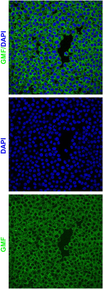
Figure 1: GMF is a novel AD therapeutic target for CRISPR-Cas9 based gene editing:
GMF immunostaining in the BV2 microglial cells indicates high-level perinuclear expression of GMF (Green) within the cytoplasm as well as on the cell surface. The nuclei (blue) are stained with DAPI. The images were acquired using the Leica TCP SP8 confocal microscope and processed using Leica Application Suite X software.

