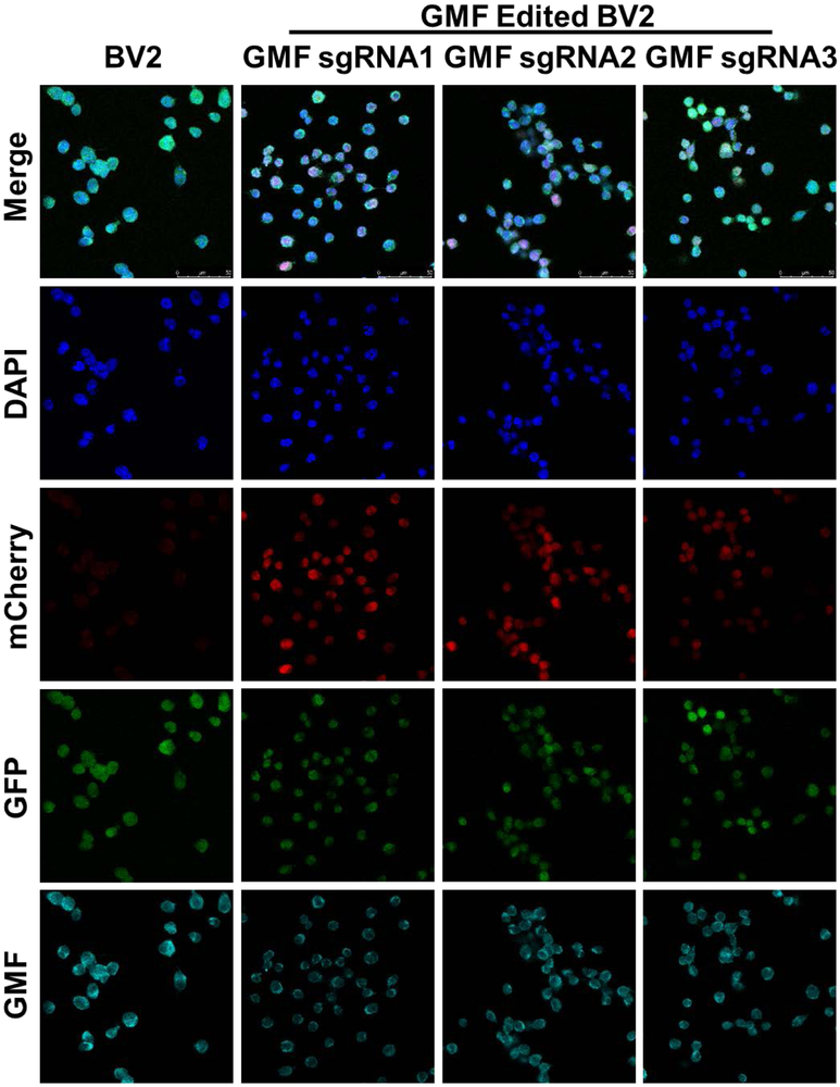
Figure 4: Targeted GMF gene editing using LV-EF1α-SpCas9-eGFP+LV-GMF-sgRNAs:
a) Confocal microscopy of the BV2-CRISPR-Cas9 cells expressing SpCas9 (Green) reveal normal GMF expression (Cyan) with DAPI stained nuclei (Blue) in the far left vertical panel labeled BV2. b) Co-transduction of BV2-CRISPR-Cas9 cells with GMF-specific sgRNA1 leads to a differential GMF editing and partial reduction in GMF expression in the vertical panel labeled as GMF sgRNA1. c) Co-transduction of BV2-CRISPR-Cas9 cells with GMF-specific sgRNA2 leads to a differential GMF editing and partial reduction in GMF expression in the vertical panel labeled GMF sgRNA2. d) Co-transduction of BV2-CRISPR-Cas9 cells with GMF-specific sgRNA3 leads to a differential GMF editing and partial reduction in GMF expression in the vertical panel labeled as GMF sgRNA3. In the panels depicting GMF sgRNAs 1-3, mCherry expression indicates co-expression of GMF sgRNAs. The GMF gene edited BV2 cells with reduced GMF expression are marked with white filled triangles.

