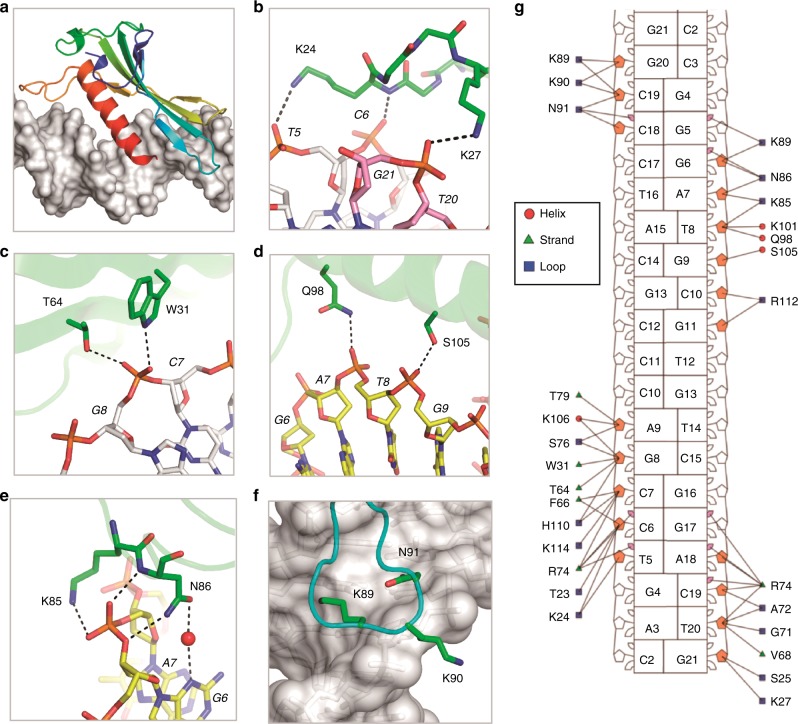
Fig. 3.
The interaction between DNA and AsfvLIG NTD. a Cartoon-and-surface view showing the relative orientation between AsfvLIG NTD and DNA. b–f Detailed interactions between AsfvLIG NTD and DNA residues. AsfvLIG NTD residues are shown as sticks in atomic colors (C, green; N, blue; O, red); the DNAs are also shown as sticks, the C-atoms of the template strand, upstream and downstream of the broken strand are colored in white, yellow, and pink, respectively. g Nucleotide-residue contact map showing individual nucleotide-residues interactions for the preferred binding site. Small and large markers on each nucleotide represent the major and minor groove contacts, respectively. Filled-in pink markers highlight which nucleotides are contacted by at least one residue in the minor groove

