Figure 7.
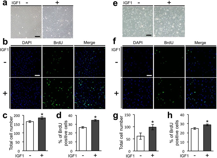
IGF1 directly upregulates the proliferation of mesenchymal and epithelial cells derived from tooth germs. (a) Phase-contrast images of mesenchymal cells isolated from mandibular molar tooth germs of ED14.5 mice at day 3 of culture. Scale bar, 200 µm. (b) BrdU-positive dental mesenchymal cells (green) were analysed by immunofluorescence. Nuclei were stained with DAPI (blue). Scale bar, 100 µm. Total numbers of dental mesenchymal cells (c) and percentages of BrdU-positive cells (d) were analysed on the immunofluorescent images. Error bars indicate the standard deviation (N = 3). *p < 0.05 (versus control group; Student’s t-test). (e) Phase-contrast images of epithelial cells isolated from mandibular molar tooth germs of ED14.5 mice at day 2 of culture. Scale bar, 200 µm. (f) BrdU-positive dental epithelial cells (green) were analysed by immunofluorescence. Nuclei were stained with DAPI (blue). Scale bar, 100 µm. The total numbers of dental epithelial cells (g) and percentages of BrdU-positive cells (h) were analysed on the immunofluorescent images. Error bars indicate the standard deviation (N = 3). *p < 0.05 (versus control group; Student’s t-test).

