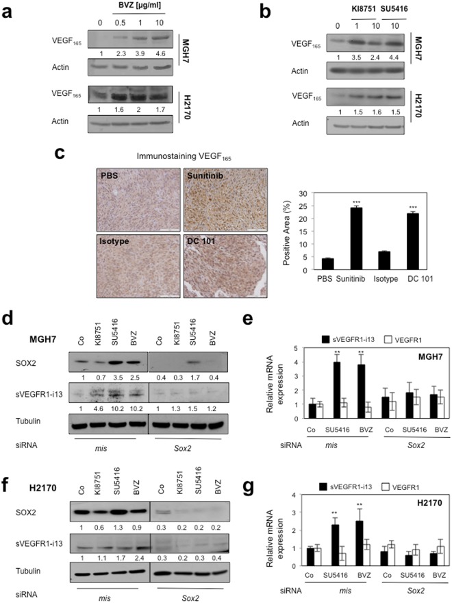
Figure 4.
Anti-angiogenic therapies up-regulate sVEGFR1-i13 expression levels in SQLC cells through a SOX2-dependent mechanism. (a,b) MGH7 and H2170 cells were treated with the indicated concentrations of bevacizumab (µg/ml) for 72 hours (a) or KI8751 or SU5416 for 24 hours (b). Western blot experiments for the detection of VEGF165. Tubulin was used as a loading control. (c) Murine SQLC tumorgrafts having received sunitinib or the murine anti-VEGFR2 antibody DC101 or not (PBS, Isotype) were recovered from previous experiments4,40. Immunostaining of VEGF165 was performed. Right panels: automatic quantification of tumor cell immunostaining for each condition. Mean ± SD of 6 mice per condition. (d,f) Western blot experiments for the detection of the indicated proteins in MGH7 (d) or H2170 (f) cells transfected during 72 hours with either mismatch or SOX2 siRNA and treated or not (Co) with 10 µg/ml bevacizumab, or transfected during 48 hours with either mismatch or SOX2 siRNA and treated for 24 additional hours with 10 µM KI8751 or 10 µM SU5416. Tubulin was used as a loading control. Black delineations allow to separate differential parts of the same gel. Quantification and statistical analyses as described above. All western blot experiments were performed at least three times. Illustrations of a representative result are presented for each condition. (e,g) RT-qPCR analyses of sVEGFR1-i13 or VEGFR1 mRNA level were performed in cells treated in the same conditions as in d or f, respectively. GAPDH was used as an internal control. The value 1 was arbitrarily assigned to the untreated condition signal.

