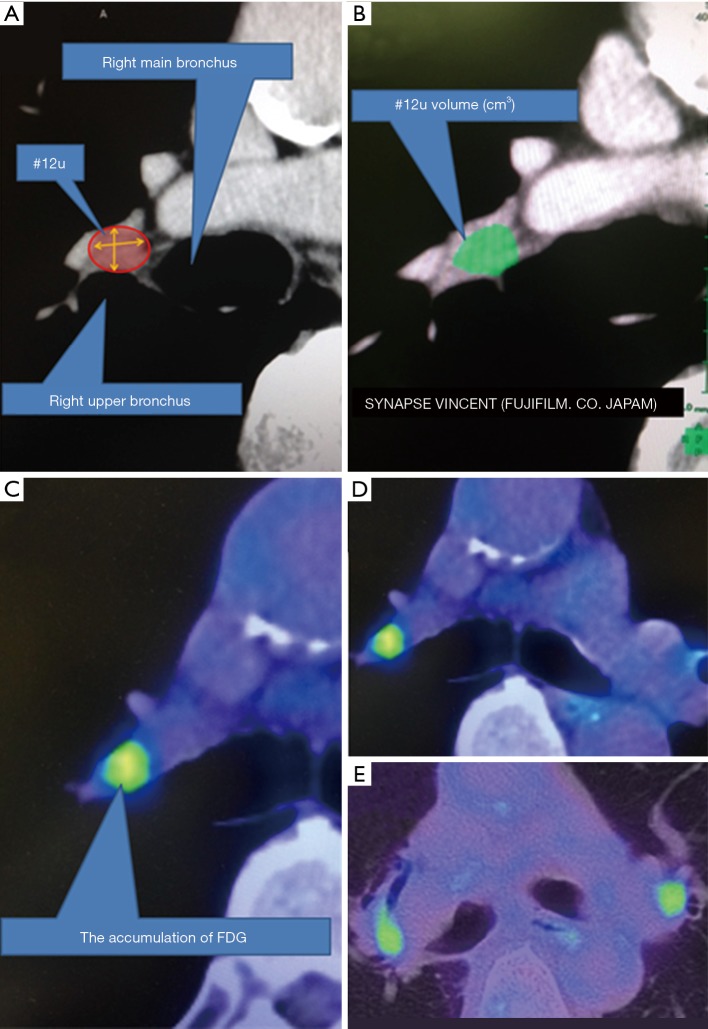
Figure 1.
Radiological image analysis. (A) #12u was defined as adjacent to the right upper lobar bronchus. It is surrounded by right main bronchus, right upper lobar bronchus, A2b of pulmonary artery branch, and right main pulmonary artery; (B) volume of #12u lymph node was measured by Synapse Vincent software; (C) SUV was measured by PET-CT. It was used for calculation of each parameter; (D) a typical image of asymmetric uptake and (E) symmetric uptake. FDG, 18F-2-floro-2-deoxyglucoce; SUV, standardized uptake value; PET-CT, positron emission tomography/computed tomography.

