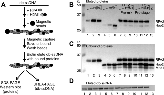
Figure 5.
RPA outcompetes Hop2-Mnd1 for binding to ssDNA. (A) Scheme for bead ‘catch and release’ of protein complexes bound to ssDNA. db-ssDNA stands for desthiobiotin modified ssDNA. (B) The captured protein complexes on ssDNA were analyzed by Western blotting. RPA were 0.125, 0.25 and 0.5 μM in lanes 1–3; 0.5 μM in lanes 7–13. Hop2-Mnd1 were 0.2, 0.4 and 0.6 μM in lanes 4–6, lanes 7–9, and lanes 10–12. Lane 13 is a control that lacked db-ssDNA but contained 0.5 μM RPA and 0.4 μM Hop2-Mnd1. (C) The unbound protein complexes analyzed by western blotting. (D) The eluted db-ssDNA–protein complexes were denatured and analyzed for DNA content on 8% urea-PAGE via staining with SYBR-gold.

