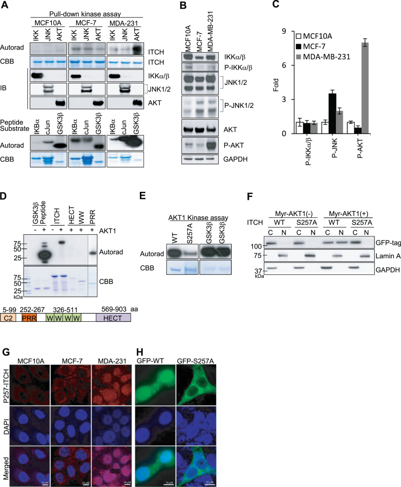
Figure 2.
Phosphorylation of ITCH at Ser257 by AKT1 is essential for nuclear translocation in TNBC. (A) In vitro kinase assay using indicated recombinant proteins as substrates for phosphorylation, visualized by autoradiography (Autorad) and Coomassie Brilliant Blue (CBB) staining for loading controls. (B) IB of indicated cell lysates stained with the indicated antibodies and (C) Quantification of fold-change relative to expression in MCF10A cells. (D and E) In vitro AKT1 kinase assays using (D) full-length and truncated ITCH proteins (as shown in schematic; not to scale) and (E) WT and S257A ITCH recombinant proteins as substrates. (F) IB of cytoplasmic (C) and nuclear (N) compartments in HEK293T cells after transient co-transfection with GFP-WT or GFP-S257 ITCH and Myr-AKT1; GAPDH and Lamin A are C and N controls, respectively. (G–H) Representative IF images of (G) indicated cell lines stained with anti-P-S257 ITCH antibody (red) and DAPI, and (H) MDA-MB-231 cells transiently transfected with GFP-WT or GFP-S257A ITCH stained with anti-GFP antibody (green) and DAPI (blue). N = 3 for IF images.

