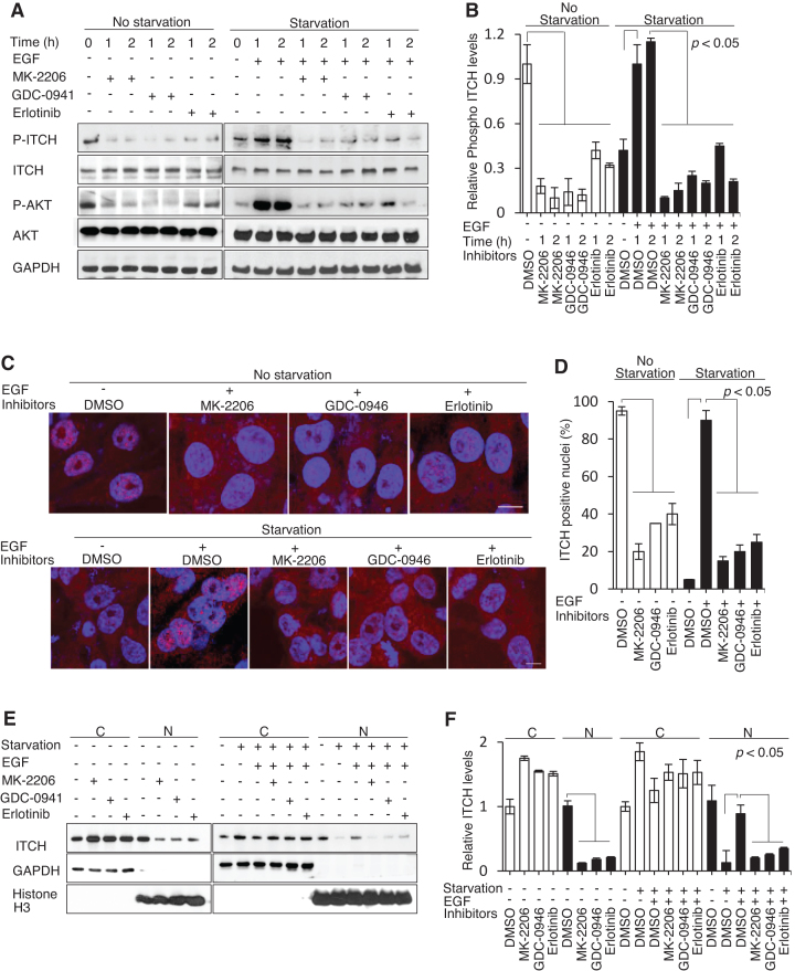
Figure 3.
ITCH nuclear translocation is mediated through an EGF/PI3K/AKT-dependent pathway. (A and B) IB using the indicated antibodies and (C and D) IF imaging using anti-ITCH antibody (red) and DAPI counterstain (blue) of MDA-MB-231 cells before and after 12–18 h 1% serum starvation with or without prior treatment with MK-2206 (0.1 μM), GDC-0941 (0.05 μM), or Erlotinib (1 μM) following the addition of EGF (10 ng/ml). (E and F) IB of cytoplasmic (C) and nuclear (N) cellular compartments. N = 3. The P values indicate significant differences between the experimental groups treated with different inhibitors (MK-2206, GDC-0946, or Erlotinib) and the control groups treated with DMSO under normal or serum-starvation conditions before and after the addition of EGF.

