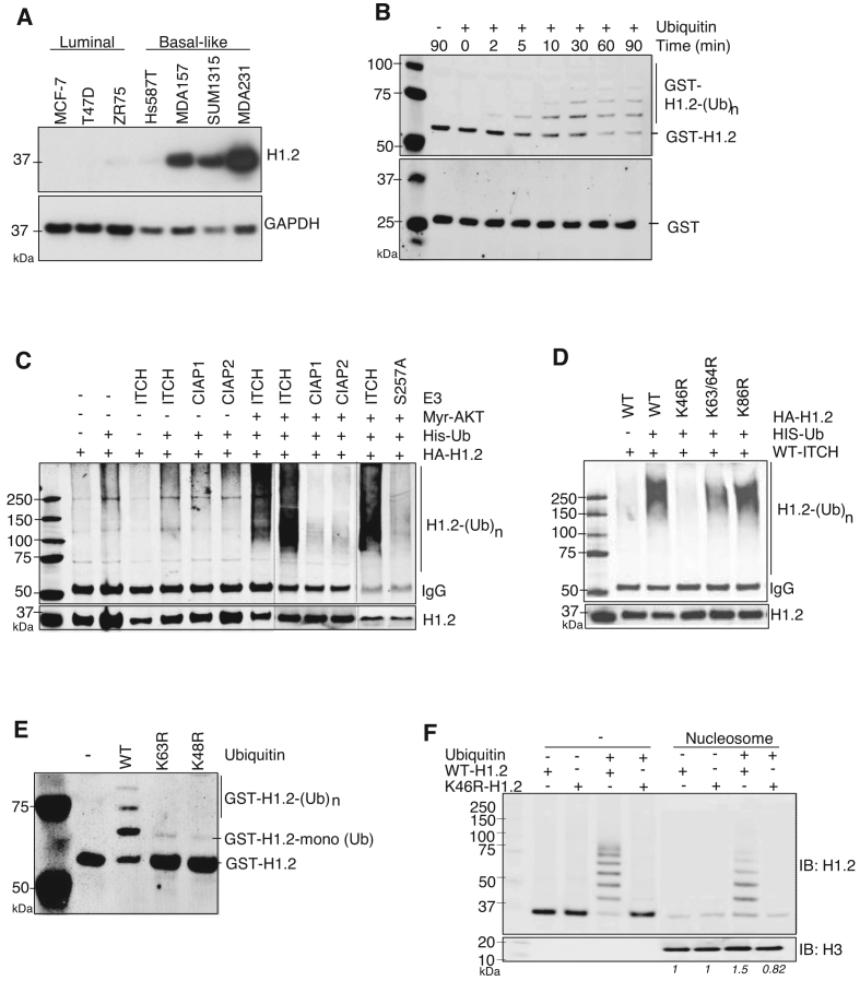
Figure 4.
ITCH interacts with and ubiquitinates linker histone H1.2 at K46. (A) IB of indicated BC cells stained with anti-H1.2 antibodies; GAPDH included as a loading control. (B) In vitro ITCH-mediated Ubn of bacterially-expressed recombinant GST alone (bottom) or GST-H1.2 (top) in the absence or presence of ubiquitin. The reaction was terminated at the indicated times and analyzed by IB using anti-GST antibody. (C and D) In vivo Ubn of H1.2 in HEK293T cells co-transfected with the indicated plasmids. Ubiquitinated HA-tagged H1.2 was IP with anti-HA antibodies followed by IB with anti-His antibodies for His-tagged Ubn; WT-, K46R-, K63/64R- or K86R-H1.2 mutants. (E) IB using anti-H1.2 antibody to measure in vitro ITCH-mediated Ubn of GST-H1.2 with WT or Ub mutants in which lysine is mutated to arginine at K63 or K48. N = 3. (F) Pull-down of recombinant HA-WT or HA-K46R H1.2 proteins –/+ Ub by biotinylated nucleosomes; IB with indicated antibodies. Relative levels of WT and K46R H1.2 binding to nucleosomes before and after ubiquitination by ITCH, normalized to the pulled down H3 protein levels, are indicated in italics.

