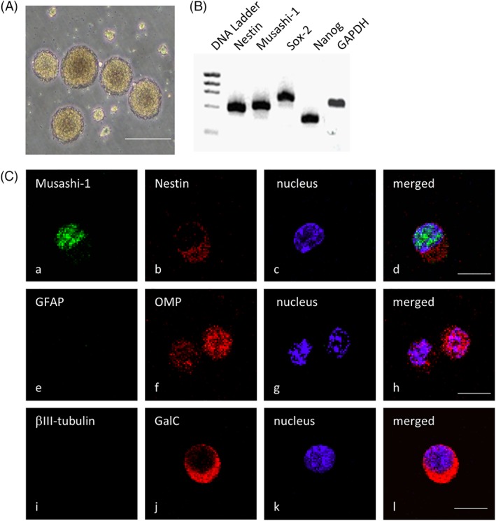
Figure 1.

Olfactory spheres and olfactory stem cells express neural stem cell markers. (A): Representative image of the floating olfactory spheres observed under a phase‐contrast microscope. (B): Reverse Transcriptase‐Polymerase Chain Reaction analysis of nestin, Musashi‐1, Sox‐2, and Nanog mRNA expression in the olfactory spheres. (C): Representative immunostaining images showing the protein expression of the neural stem cell markers Musashi‐1 (Ca), nestin (Cb), and OMP (Cf) as well as the expression of GFAP (Ce), βIII‐tubulin (Ci), and GalC (Cj), which label differentiated astrocytes, neurons, and oligodendrocytes, respectively. The nuclei were counterstained with Hoechst 33258 (Cc, Cg, and Ck), and merged images are shown in (Cd), (Ch), and (Cl). The scale bars indicate 100 μm (Ca) and 50 μm (Cc).
