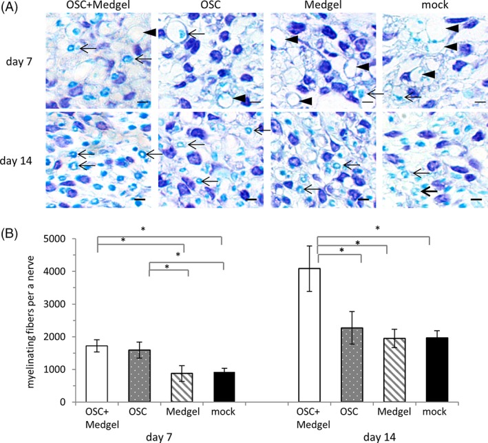
Figure 6.

Stimulation of histopathological recovery by olfactory stem cells with Medgel. (A): Magnified images of sections of the right facial nerves 1 mm distal to the compression site 7 days (upper row) or 14 days (lower row) post‐treatment after staining with the Klüver–Barrera method. Arrowhead indicates nerve fibers with degraded myelin, whereas the arrows highlight regenerating nerve fibers with intact myelin. The scale bars indicate 100 μm. (B): The numbers of myelinated nerve fibers calculated for each group (n = 3). The data are presented as the means ± standard deviation. * p < .05.
