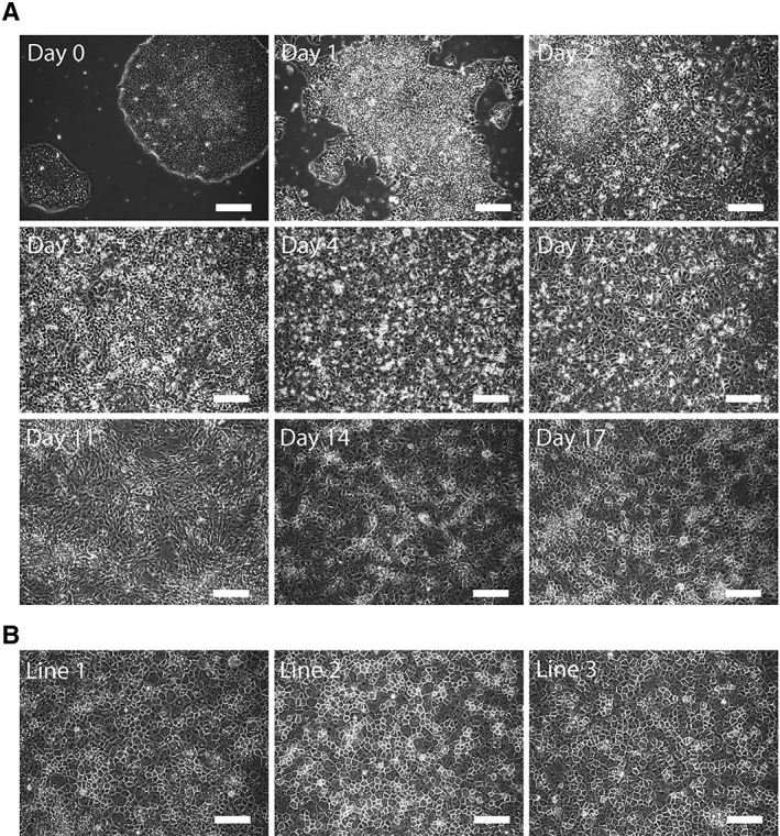
Figure 2.

Morphological characterization of hepatic differentiated cells. (A): Brightfield microscopy images revealing the morphological transformation from day 0 pluripotent stem cell colony to day 17 polyhedral hepatocytes. (B): Representative images of day 21 human pluripotent stem cells‐Heps generated from three different current good manufacturing practice‐compliant lines. Scale bars: 100 μm.
