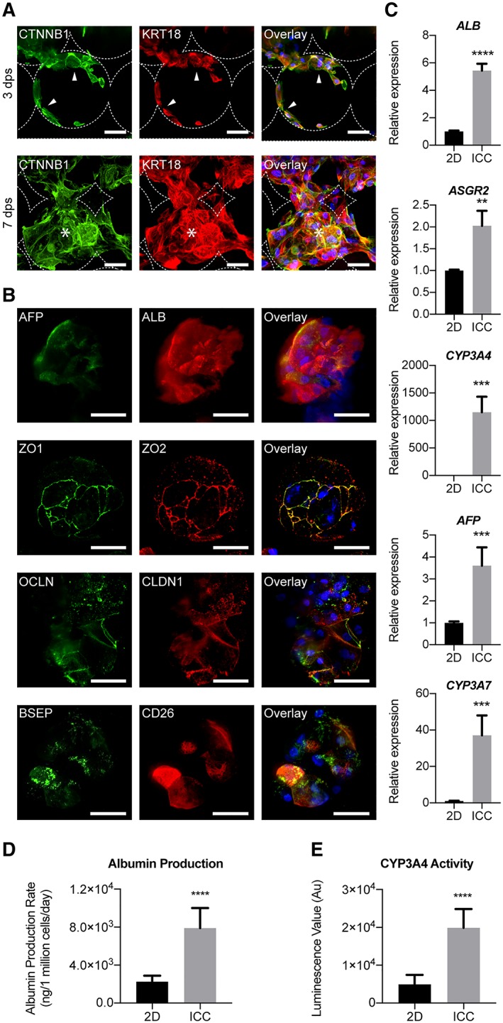
Figure 5.

Characterization of human pluripotent stem cells (hPSC)‐derived hepatocyte maturation within three‐dimensional (3D) inverse colloidal crystal (ICC) model. (A): Immunofluorescent confocal images of hPSC‐Heps demonstrating two distinguished morphological phases inside the ICC scaffold. Three days postseeding an adhered lining across the hydrogel pores is observed, before the hPSC‐Heps morph into interconnected 3D clusters from 7 days postseeding onward. Arrowheads indicate cells lining the ICC scaffold surface; asterisks represent cells forming 3D clusters. Scale bar, 100 μm. (B): Immunofluorescent confocal images highlighting hepatic (AFP and ALB) and polarity (ZO‐1, ZO‐2, OCLN, CLDN1, BSEP, and CD26) proteins known to be present in adult human hepatocytes. Scale bar, 100 μm. Staining was performed on cell clusters after hPSC‐Heps had been cultured for 2 weeks in 3D. (C): Real‐time polymerase chain reaction showing the relative expression of five major hepatic genes (ALB, ASGR2, CYP3A4, AFP, and CYP3A7); n = 4 experiments, one cell line. (D): Albumin production rate of hPSC‐Heps cultured in 2D versus ICC models; n = 4 experiments, one cell line. (E): CYP3A4 basal activity of hPSC‐Heps cultured in 2D versus ICC scaffolds; n = 4 experiments, one cell line. Data are mean ± SD. Student's t test (two‐tailed) analysis. **, p < .005; ***, p < .0005; ****, p < .0001. Data shown for cell line 1.
