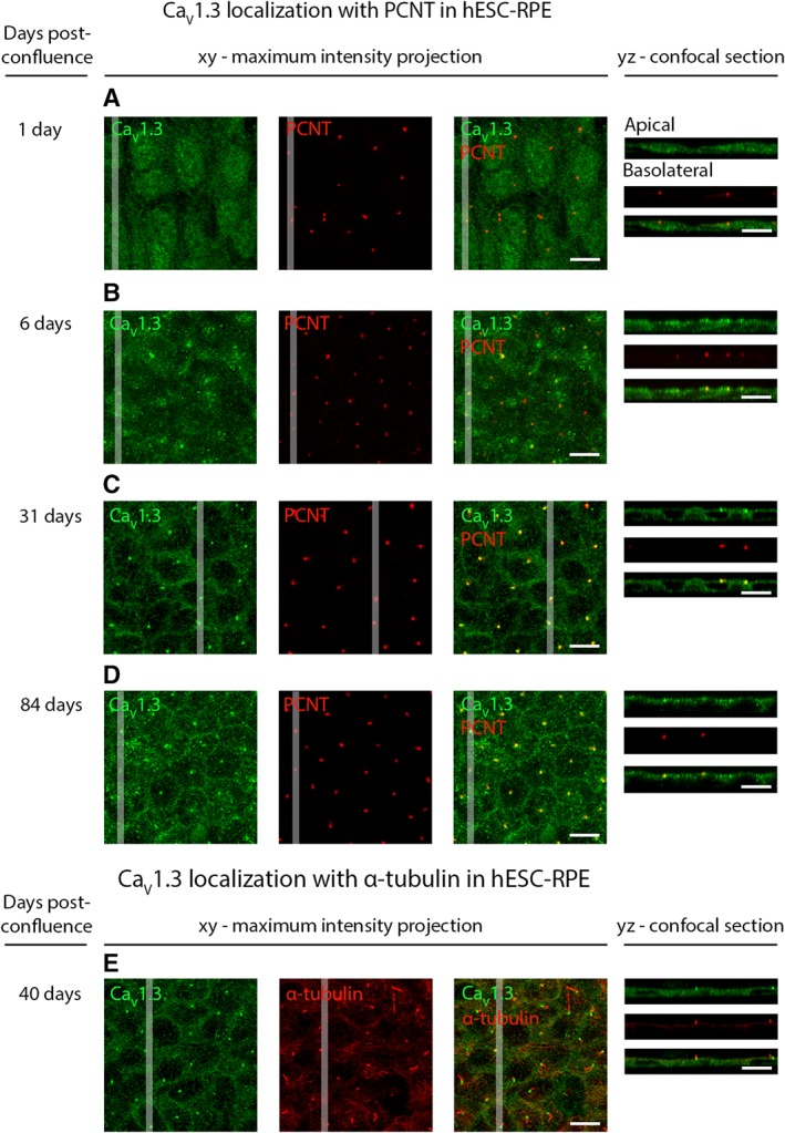
Figure 7.

Localization of CaV1.3 during hESC‐RPE maturation. Immunolabeling of CaV1.3 (green) together with centrosome protein PCNT (red) from post‐confluence day 1 to post‐confluence day 84 at four time points: (A) day 1, (B) day 6, (C) day 31, and (D) day 84 (cell line 08/017). (E): Labeling acetylated α‐tubulin (red) together with CaV1.3 (green) shows the localization of CaV1.3 at the base of the primary cilia during maturation (cell line 08/017, days post‐confluence 40). The confocal images are shown as xy‐maximum intensity projections and yz‐confocal sections (apical side upwards, localization of the section highlighted with a white bar). Scale bars 10 μm. Abbreviations: CaV, voltage‐gated Ca2+ channel; hESC, human embryonic stem cell; RPE, retinal pigment epithelium; PCNT, pericentrin.
