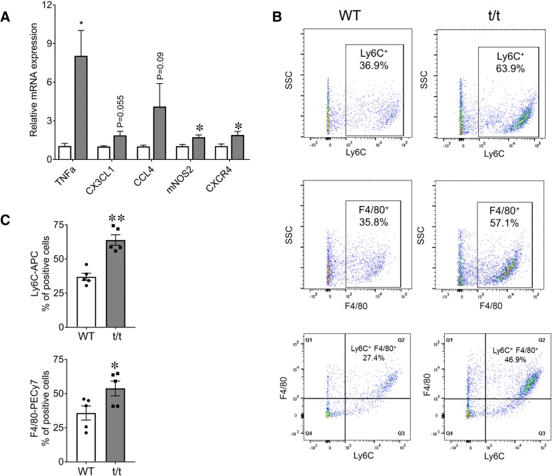
Fig. 5.
Elevated expression of inflammatory markers and cells in t/t GAS. a mRNA levels of inflammatory mediators and chemokine receptors measured by RT-qPCR in GAS muscles showed increased expression in t/t compared to WT mice. Representative flow cytometry analysis of monocytes showed Ly6C+- and F4/80+-gated cells from GAS muscle of t/t and WT mice (b). The percentage of Ly6C+ and F4/80+ cells (c) was significantly greater in GAS of the t/t mice (Ly6C+ − 36.9% in WT vs. 63.9% in t/t mice (n = 5, **P < 0.001); F4/80+ − 35.8% in WT vs. 57.1% in t/t mice (n = 4, *P < 0.02)). Ly6C+ cells were further sorted by F4/80 gating to determine if the tissue-specific macrophages were positive for the inflammatory marker. t/t mice showed an increase in the double positive population (as shown in Q2), indicating a greater number of intramuscular inflammatory macrophages (b, bottom)

