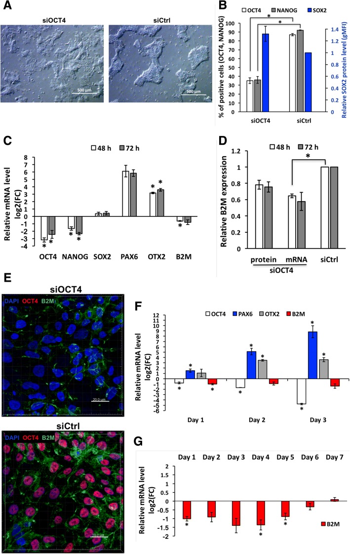
Fig. 4.
Expression of B2M is downregulated during early neuroectodermal differentiation induced by OCT4 silencing or neural induction medium. a Representative light micrographs illustrating changes in hES cell morphology at 72 h caused by treatment with 20 nM siOCT4. Scale bar is 500 μm. b Flow cytometric analysis of core pluripotency factor expression at protein level at 72 h in cells treated with 20 nM siOCT4, represented as percentage of cells positive for OCT4 and NANOG, and as a protein level (estimated according to gMFI change) normalized to siCtrl for SOX2. For gating strategy, see the “Methods” section. c RT-qPCR analysis of representative pluripotency and differentiation markers expression upon treatment with 20 nM siOCT4. mRNA level is presented as logarithm base 2 of the fold change in gene expression between the siCtrl and siOCT4 sample with identical concentration. d Flow cytometry and RT-qPCR analysis of B2M expression upon treatment with siOCT4. Values represented are relative to siCtrl sample with identical concentration (20 nM). e Representative fluorescence micrographs of OCT4 (red) and B2M (green) expression at 72 h in samples treated with 20 nM siOCT4 or siCtrl. Cell nuclei were stained with DAPI. Scale bar is 20 μm. f–g RT-qPCR analysis of representative pluripotency and differentiation markers and B2M expression during growth in neural induction medium. mRNA level is presented as a logarithm base 2 of the fold change in gene expression between cells grown in mTeSR1 and neural induction medium. Abbreviations: FC, fold change. Statistical significance with P values less than 0.05 are labeled as * (mean ± SEM, N = 3)

