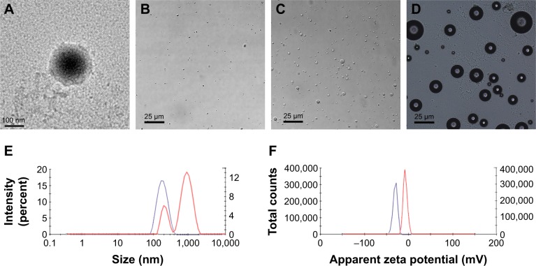
Figure 1.
Characterization of TOI_HNPs.
Notes: (A) TEM image of TOI_HNPs. (B, C) Optical microscopy images of TOI_HNPs and phase-transited TOI_HNPs after laser irradiation. (D) Optical microscopy image of TOI_HNPs heated at 49°C for 10 seconds. (E, F) Size distribution and zeta potential of TOI_HNPs before (the blue line) and after (the red line) laser irradiation measured by DLS.
Abbreviations: DLS, dynamic light scattering; ICG, indocyanine green; PFP, perfluoropentane; LPHNPs, lipid–polymer hybrid nanoparticles; TEM, transmission electron microscope; TOI_HNPs, folate-targeted LPHNPs-loaded ICG/PFP-carrying oxygen.

