Figure 2.
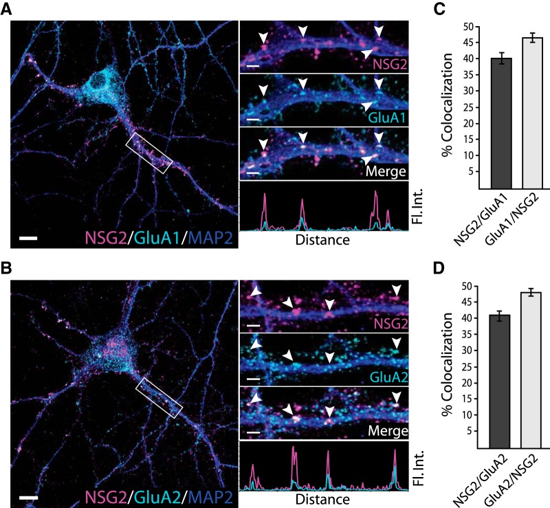
NSG2 colocalizes with AMPAR subunits GluA1 and GluA2. A, B, Representative confocal images of primary hippocampal neurons at DIV15 showing perinuclear and punctate NSG2 (magenta) in MAP2+ (blue) neurites along with surface expressed GluA1 punctae (A, cyan), and GluA2 punctae (B, cyan). Separated color panels for individual markers from the boxed regions have been magnified for clarity (right panels). Lower right panels in A, B show the colocalization profile for NSG2 (magenta)/GluA1 (cyan, panel A) and NSG2 (magenta)/GluA2 (cyan, panel B) across the indicated punctae (arrowheads). C, D, Quantification of colocalization for NSG2 with surface GluA1 punctae (6423 NSG2 and 5392 GluA1 puncta from 10 neurons) and NSG2 with surface GluA2 punctae (6874 NSG2 and 5963 GluA2 puncta from nine neurons), respectively, from three independent cultures. Scale bars = 10 μm (A, B; left panels) and 2 μm (A, B; right panels).

