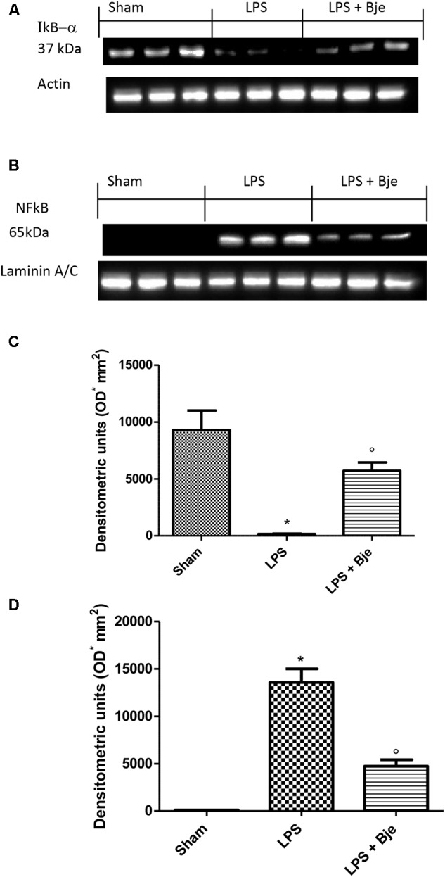
FIGURE 4.

BJe inhibits NF-κB activation. Blot in (A) and its densitometric analysis in (C) showed that BJe significantly decreased IκB-α degradation (A,C). Blot (B) displayed that LPS injection increased NF-κB translocation in the nucleus (B,D), whereas BJe significantly reduced the presence of NF-κB in the nuclear fraction (B,D). Levels of IκB-α and NF-κB presented in the densitometric analyses of protein bands were normalized for β-actin and laminin, respectively. Data reported are presented as mean ± SEM (N = 10 rats for each group). ∗P < 0.05 vs sham group. °P < 0.05 vs LPS group.
