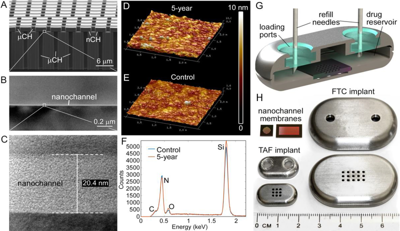
Figure 1. Nanochannel membrane and NDI.
(A) Scanning electron microscopic image of nanochannel (nCH) membrane with microchannels (μCH). (B), (C) Transmission electron microscopic image depicting 20 nm nanochannel. (D) Representative AFM image of membrane in PBS at 23 °C over 5 years compared to (E) fresh membrane. (F) EDX analysis of surface elements below TaN/SiC coating of membrane in PBS at 23 °C over 5 years compared to fresh control. (G) Cross-sectional rendering of NDI depicting drug refill needles through the loading ports with resealable silicon plugs. (H) Nanochannel membranes of two different sizes, square (TAF) and rectangle (FTC); top and bottom view of NDI in medical-grade titanium for TAF (with silicon plug) and FTC (before affixing silicon plug).

