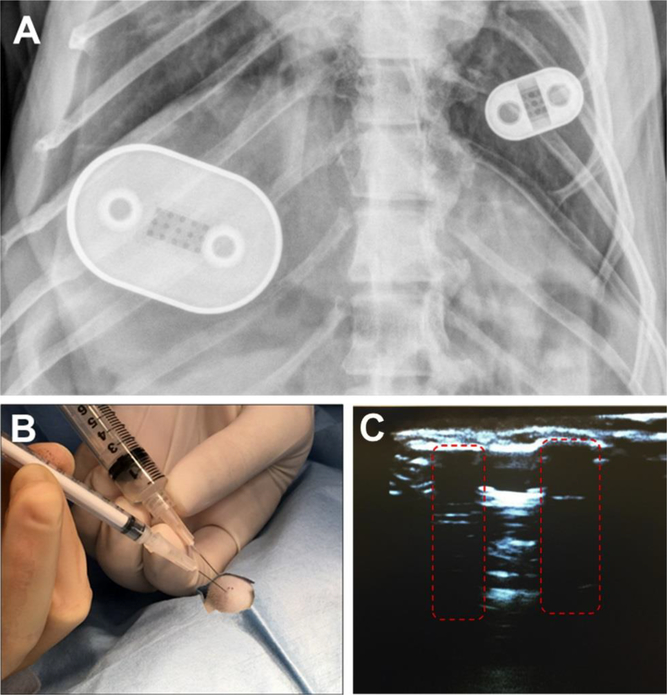
Figure 6. NDI implants in rhesus macaque on day 49 and transcutaneous drug refilling.
(A) X-ray radiography image on day 49 of NDI-FTC (left) and NDI-TAF (right) implanted subcutaneously in the dorsum of a rhesus macaque. During surgical implantation of the NDIs, the side with drug refilling ports was positioned up and drug-eluting side with nanochannel membrane facing down. (B) Transcutaneous refilling procedure of NDI-TAF in the dorsum of a rhesus macaque. A venting syringe was inserted into one port, while a drug solution-filled syringe was inserted into the other port. Injection of drug was performed simultaneous to the withdrawal of residual fluid through the venting syringe. (C) Ultrasound image of loading and venting ports (red dashed boxes) of NDI- FTC subcutaneously implanted in a pig cadaver.

