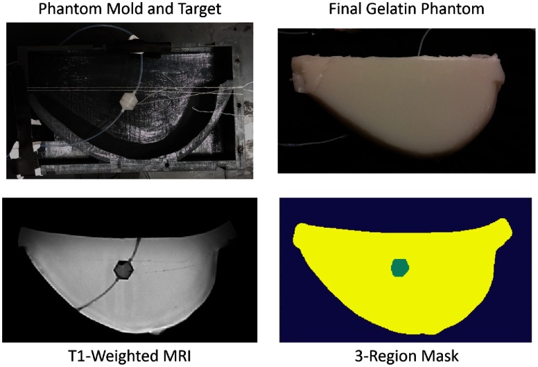
Fig. 5.
Gelatin phantom and mold. (a) One half of the breast-shaped gelatin phantom mold with target, prior to pouring of the gelatin mixture. The target is held in place by thin, white string which does not significantly change the optical properties of the phantom. (b) Final gelatin phantom. Different liquid phantom materials can be injected, via the nylon tubing, into the target within the gelatin. (c) T1-weighted MR image of the phantom. (d) Signal-threshold segmentation of MR image to define distinct regions in the tissue phantom. Here, the image is segmented into a three region-types, i.e., the liquid target, the gelatin phantom, and the background region. A three-region segmented image mask is then ready for use as a hard- or soft-prior constraint for DOT.

