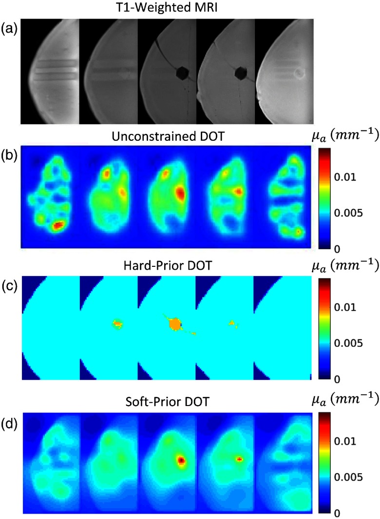
Fig. 6.
Unconstrained, hard-prior, and soft-prior DOT reconstructions. (a) Sagittal slices, separated by 1 cm, of a T1-weighted fat-suppressed gradient echo MRI of the gelatin phantom and target. Note that the horizontal lines on the first (source-side) and last (detector-side) slices are indentations in the phantom from the optical modules. (b) Sagittal slices of an unconstrained DOT reconstruction. The DOT reconstruction accurately localizes the target and provides good absorption contrast relative to the background. However, numerous boundary artifacts also arise with reconstructed values similar to the targets. (c) Sagittal slices of a DOT reconstruction constrained by a hard spatial prior. Interestingly, although the artifacts are removed by the hard-prior constraint, the contrast here is also reduced. This effect could be due to slight inaccuracies in the co-registration of the segmented MR image. (d) Sagittal slices of a DOT reconstruction constrained by a soft spatial prior. Notice that the contrast is slightly improved and the artifacts are significantly reduced relative to the unconstrained reconstruction.

