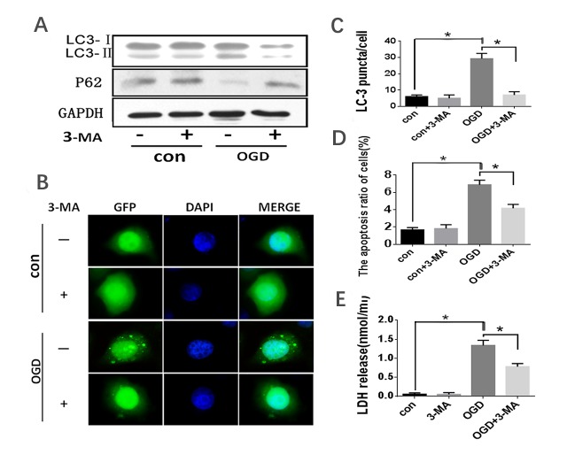
Figure 3.
OGD treatment induced autophagy activation in HL-7702 cells. HL-7702 cells underwent OGD for 40 minutes with or without pre-treatment of 5 mM 3-MA for 3 hours. (A) Western blot analysis with the indicated antibody (LC3 and p62 antibody for detecting autophagy; GAPDH was used as a loading control). (B) Confocal microscopy was used to detect the formation of GFP-LC3 puncta. Original magnification, 400×. (C) Quantification of cells with >5 GFP-LC3 puncta. The data were presented as the mean ±SD from three independent experiments. (D) Apoptosis was assessed by flow cytometry with Annexin V/PI stain. (E) Cell death was assessed by LDH release into the supernatant. *p<0.05.

