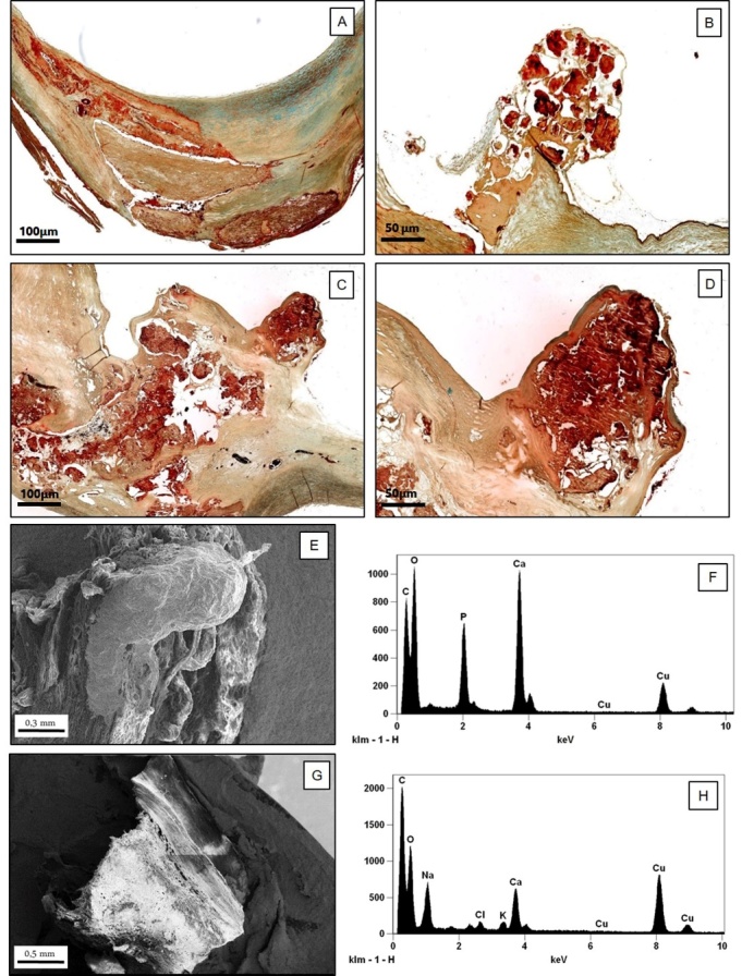
Figure 1.

Characterization of nodular calcification. A-D) Histological and ultrastructural characterization of calcifications in carotid plaques. (A) Stable fibrocalcific plaque with a linear calcification (Movat staining,2x); (B) Calcific carotid nodule protruding into the lumen consisting of calcified plates associated to a small amount of fibrous tissue without extracellular lipids, necrotic core and inflammatory cells. The fibrous cap over the nodule was extremely thin (Movat staining, 5x); (C, D) Images display a calcific nodule associated to fibrin deposition with a small superficial ulceration (C): Movat staining, 2x, D (particular of C): 6x). (E-H) Scanning electron microscopy and EDX microanalysis of carotid plaque: (E) The image shows an eruptive calcific nodule with disruption of fibrotic cap (scanning electron microscopy, 100x); (F) EDX spectrum shows that this calcific nodule is made from hydroxyapatite; (G) Linear calcification in a stable fibrocalcific carotid plaque (scanning electron microscopy, 90x); (H) EDX spectrum demonstrates that this linear calcification is made from calcium oxalate.
