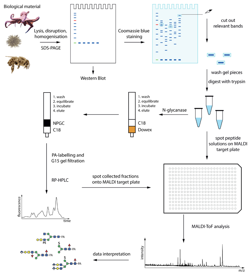
Figure 1. A potential glycome and glycoproteomic workflow.
Starting from biological material, proteins can be separated by SDS-PAGE prior to Western blotting or peptide map fingerprinting. The peptides and glycopeptides are analysed directly by mass spectrometry; the glycans are released by an N-glycanase such as PNGase Ar and purified by two rounds of solid phase extraction prior to mass spectrometry and/or HPLC. Glycans (examples from honeybee royal jelly) are depicted according to the Symbol Nomenclature for Glycans, whereby circles, squares, triangles and diamonds respectively represent hexose (here Man or Gal), N-acetylhexosamine (GalNAc or GlcNAc), deoxyhexose (Fuc) or hexuronic acids (GlcA); S, sulphate; PE, phosphoethanolamine.

