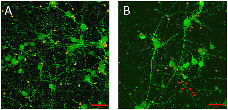
FIGURE 1 |.
Embryonic 12.5 (E12.5) hindbrain cells encapsulated in 3D hydrogel scaffolds (GelMA/HAMA: 3.5/0.5 wt%, no laminin added) remained viable after 10 days in vitro (10DIV). (A) Maximum intensity projection depicts cell bodies projecting extensive processes adopting elaborated arborization patterns. Viable cells, shown in green, were labeled with calcein-AM (see Materials and Methods section for details). Scale bar: 50 μm. (B) Some encapsulated E12.5 hindbrain cells [same conditions as (A)] showed varicose neurites. Red arrows indicate the putative boutons along the neurite outgrowth. Scale bar: 50 μm.

