
Fig 1. Morphological observation of field-collected cysts/zygotes of snow-inhabiting Chloromonas.
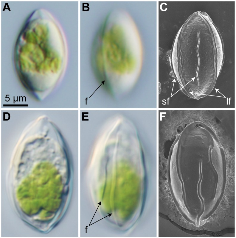
Identical magnification throughout. For detailed information of collection sites, see S1 Table. (A–C) “C. nivalis zygotes” from site 160518Hk2G1 in Mt. Hakkoda, Japan. (A, B) Light micrographs. (A) Optical section. (B) Surface view, showing a flange (f). (C) Field emission scanning electron micrograph. Abbreviations: lf, long flange (extending the entire cell length); sf, short flange (reaching neither pole of the cell). (D–F) C. miwae cysts from site 130630Gs4G in Mt. Gassan, Japan. (D, E) Light micrographs. (D) Optical section. (E) Surface view, showing flanges (f). (F) Field emission scanning electron micrograph.