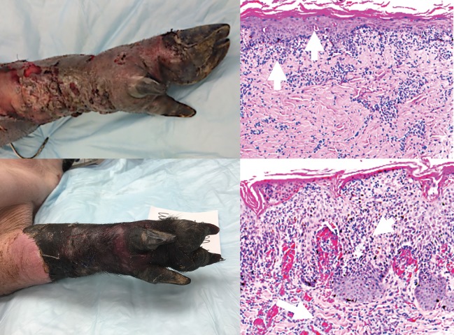
Fig 7. Clinical and histopathologic assessment of rejection.
Top left panel: Banff Grade 3 AR (clinical picture) Top right panel: Histopathology of Grade 3 AR showing dense inflammation and epidermal involvement with apoptosis, dyskeratosis, and/or keratinolysis (arrows). Bottom left panel: Banff Grade 4 AR (clinical picture) Bottom right panel: Histopathology of Grade 4 AR showing necrotizing acute rejection. Frank necrosis of epidermis and presence of microvascular thrombi in deep dermal capillaries.

