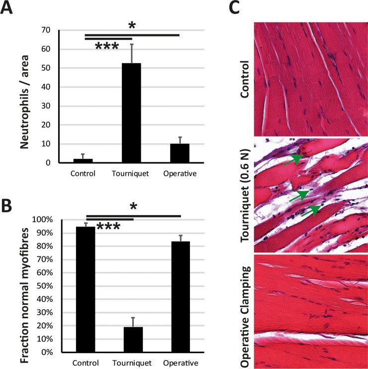
Fig 5. Low-pressure tourniquet leads to destruction of muscle fibers and neutrophil infiltration.
After ischemia of 2 hours and reperfusion of 4 hours (A) Neutrophils per 2000×2000 Pixels were measured. The low-pressure tourniquet led to a significant increase in neutrophil infiltration compared to control (sham) animals. The operative clamping of the femoral artery did not show the same significant increase in neutrophil infiltration. (B) Fraction of normal myofibers was significantly lower in the tourniquet cohort compared to control and operative clamping. (C) Histological images at a magnification of 100× showed a higher neutrophil infiltration (neutrophils are marked with a green arrow) in the tourniquet group compared to control and operative clamping. Results are shown as means ± SEM. P-value: * < 0.05; *** < 0.001; (ANOVA), n = 4.

