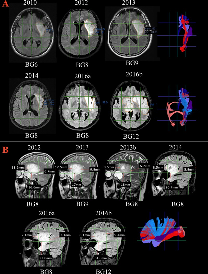
Fig 6. The radiological course of Illustrative case #2.
A) Axial slices showing morphological FLAIR MR sequences during a longitudinal follow-up from 2010 and onwards. The signal hyperintensity evolved from 6 Brain-Grid units to 12 units, with a clear morphological transformation from a bulky shape to a diffuse and digitated shape infiltrating along the subcortical white matter. On the right, tractographic reconstructions from the Brain-Grid atlas revealed the major white matter involved during the progression. External capsule (light blue), UF (violet), IFOF (red), anterior thalamic radiation (dark blue), MLF (purple), AC (salmon). Follow-up MRIs in 2012 and 2013 demonstrated increased tumor volume involving also a radial extension of hyperintensity from the insula through the extreme and external capsule. Eight grid voxels were infiltrated in 2012, 9 voxels in 2013, with involvement of the S3 areas on both the lateral side (A4) and medial side (A3) within the intermediate coronal area (C2). The invasion at this time point, as shown in A–B, is more prominent through the posterior portion of the insula and sub-insular white matter. The potential pathways of infiltration are represented by the MLF fibers on the lateral (A4) grid voxel and the IFOF fibers medially (A3), caudally through the periventricular white matter (within the C3 area). After radiotherapy, the tumor volume as well as the infiltration along the longitudinal posterior pathways decreased significantly. The number of segments decreased to 8 due to a reduction of the hyperintensity in the A3C3S3 voxel. B) Details of the radiological follow-up between 2011 and 2016 that capture the switch from a bulky shape to a more diffuse and infiltrative appearance. On the right side, the Brain-Grid tractographic reconstructions summarizing the major white matter bundles involved during tumor progression. External capsule (light blue), UF (violet), IFOF (red), anterior thalamic radiation (dark blue), MLF (purple), AC (salmon). The sagittal projection shows that further infiltration along the antero-ventral pathways (UF) was not prevented by radiotherapy and that slow but continuous tumor growth occurred during the 3 years following radiotherapy. In 2016, when the entire anterior temporo-basal area was infiltrated, the number of infiltrated grid voxels was 12, showing also interhemispheric spread through the anterior commissure to the medial (A2) and intermediate coronal (C2) S2 and S3 grid voxels.

