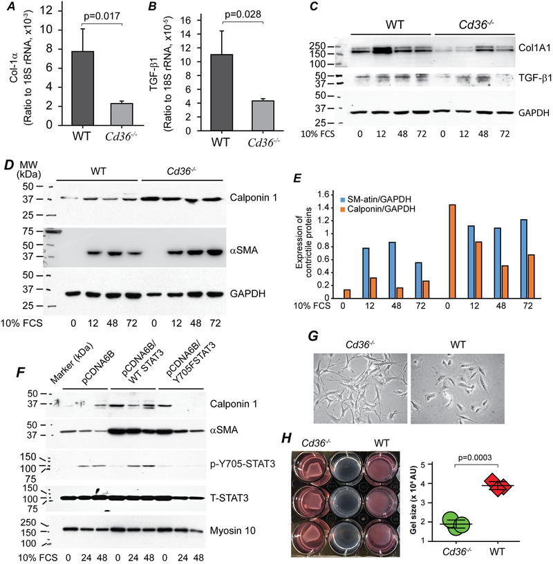
Figure 7. CD36 deficiency switches VSMCs to a contraction phenotype.
A & B. WT and Cd36−/− cells were cultured in 10 cm plates (106 cells) for 24 hrs and serum starved for 24 hrs. Cells were then stimulated with DMEM containing 10% FCS for an additional 24 hours. Total RNAs were isolated, converted to cDNA and qPCRs was performed for Col1A1 (A) and TGF-β (B), and data were normalized to 18s rRNA, n=3/group. C, D and E. Serum-starvation synchronized WT and Cd36−/− cells were stimulated with 10% FCS for the indicated times, and then Col1A1, TGF-b1 precursor (C), as well as Calponin 1 and aSMA (D & E) were assessed by Western blot assay. GAPDH served as loading control in both blots. F. Cell lysate prepared as mentioned in Fig. 6D were used for examination of Calponin 1, aSMA and myosin 10. Blots for T-STAT3 and p-Y705-STAT3 were same to that shown in Fig. 6D. G. Phase-contract images of WT and Cd36−/− cells. H. Collagen-gel based cell contraction assay.

