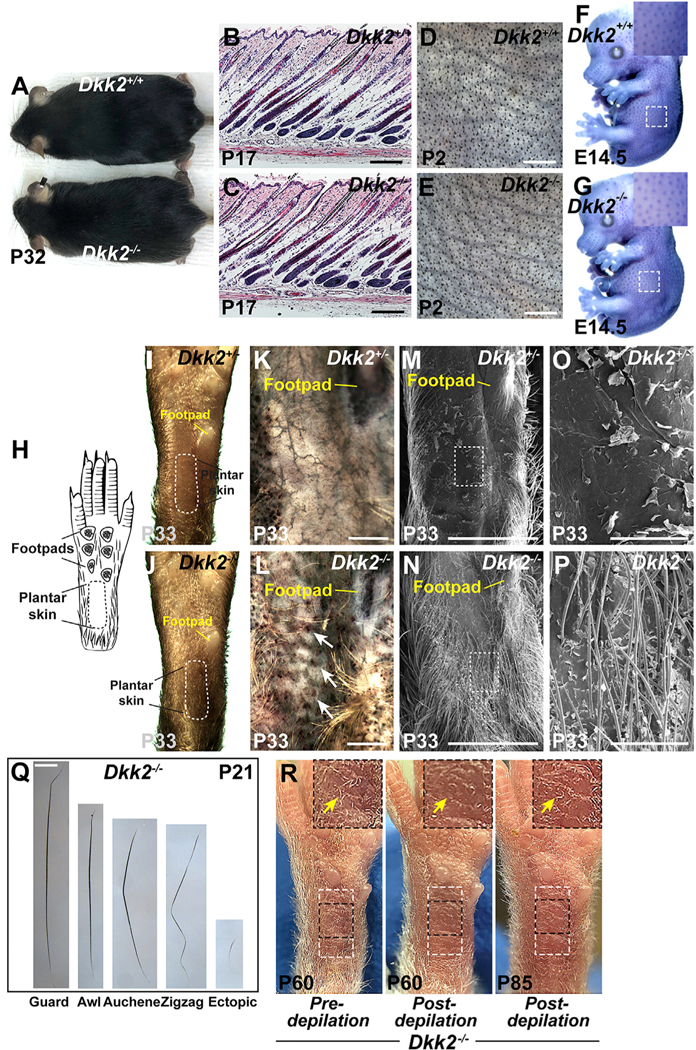
Figure 1. Ectopic Hair Follicles Form In Plantar Skin of Dkk2 Null Mice.

(A)Normal appearance of the hair coat of Dkk2−/− mouse at P32 compared with a wild-type littermate control (n > 40 mutants and littermate controls). (B and C) Indistinguishable histology of Dkk2−/− (C) and Dkk2+/+control littermate (B) dorsal skin at P17 (n = 3 mutants and n = 3 littermate controls). (D and E) Light microscopy of dissected dorsal skin at P2 reveals similar density and patterning of primary and secondary hair follicles in Dkk2−/− (E) and Dkk2+/+ control (D) littermate mice (n = 3 mutants and n = 3 littermate controls). (F and G) Whole mount in situ hybridization for Ctnnb1 (purple-blue signal) reveals similar sizes and patterning of primary hair follicle placodes in Dkk2−/− (G) and Dkk2+/+control (F) littermate embryos at E14.5 (n = 3 mutants and n = 3 littermate controls). Insets: higher magnification views of the boxed areas. (H) Schematic indicating the locations of footpads and plantar skin on the ventral side of a right hind foot. A dashed black line marks the border of plantar skin. (I-P) Light microscopy (I and J), alkaline phosphatase staining (purple/brown) of dissected plantar dermis (K and L), and scanning EM micrographs (M-P) of the ventral side of right hind feet from Dkk2+/−control (I, K, M, and O) and Dkk2−/− (J, L, N, and P) mice at P33 (n > 40 mutants and n > 40 controls for I and J; n = 3 mutants and n = 3 littermate controls for each experiment in K-P). The plantar region is delineated by a dashed white line in (I) and (J). White arrows in (L) indicate ectopic alkaline phosphatase-positive hair follicle dermal papillae. (O) and (P) are higher magnification views of the boxed regions in (M) and (N), respectively. Locations of the footpads are indicated in (I)-(N). Note formation of fully formed ectopic hair in Dkk2−/− plantar skin. (Q) Morphology of ectopic hair shaft compared with normal guard, awl, auchene, and zigzag hairs plucked from ventral limb skin at P21. Ectopic hair is shorter and finer than normal hair (n = 3 mutant mice analyzed). (R) Photographs of right hind foot of a Dkk2−/− mouse prior to depilation of the plantar region at P60 (left), immediately following depilation (middle), and 25 days later (P85) (right). Areas outlined with a dashed white box indicate the plantar region. Insets: higher magnification views of areas outlined by dashed black boxes. Arrows indicate ectopic plantar hair (left); lack of hair after depilation (middle); and re-growth of ectopic hair at P85 (right) (n = 3 Dkk2−/− mice depilated). Scale bars, 150 μm (B and C); 1.5 mm (D and E); 359 μm (K and L); 2 mm (M and N); 300 μm (O and P); 700 μm (Q). See also Figure S1.
