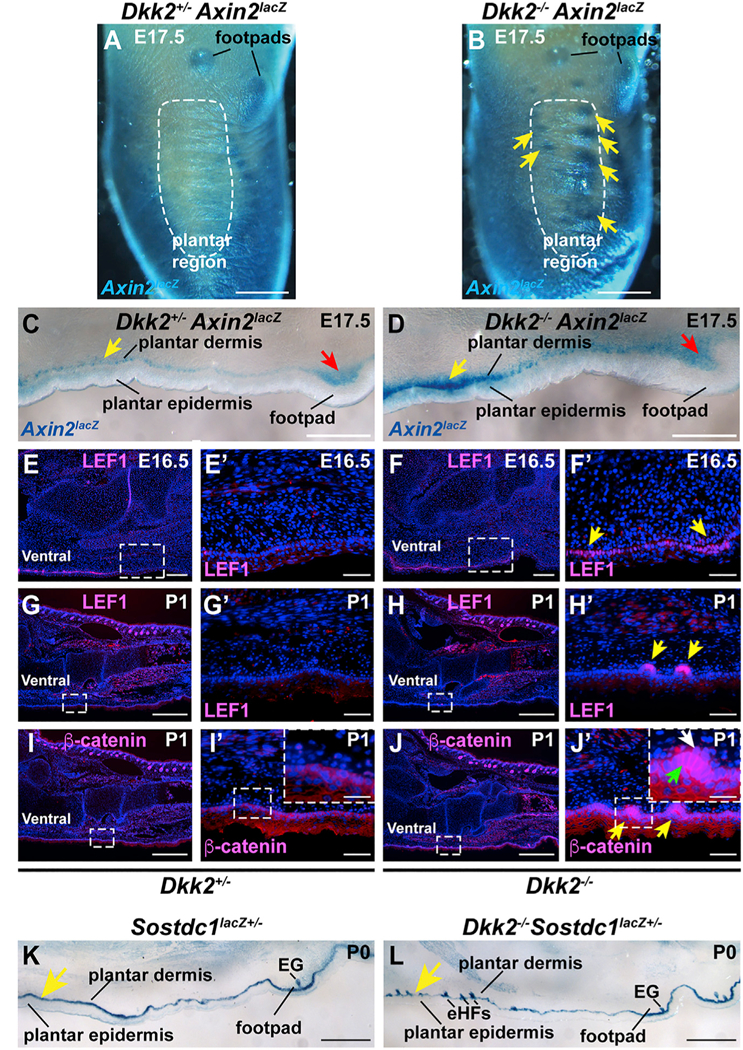
Figure 5. Wnt/β-Catenin Signaling Is Elevated in Embryonic and Neonatal Dkk2−/− Plantar Skin.

(A and B) X-gal-stained whole mounted control Dkk2+/− Axin2lacZ (A) and Dkk2−/− Axin2laaZ (B) hind paws at E17.5. Note elevated Axin2lacZ Wnt reporter activity (blue signal) in the developing plantar region of the Dkk2−/− mutant (B, arrows). The plantar region is outlined by a dashed white line in (A) and (B) and footpad locations are indicated. (C and D) Cryosections of ventral skin from X-gal stained Dkk2+/− Axin2lacZcontrol littermate (C) and Dkk2−/− Axin2lacZ (D) hind paw whole mounts at E17.5. Wnt reporter expression is elevated in plantar papillary dermis and plantar epidermis in the Dkk2−/− mutant compared with the control (yellow arrows). Signaling levels are similar in mutant and control footpads (red arrows). (E-JꞋ) Hind paw sections from Dkk2+/− control littermate (E, EꞋ, G, GꞋ, I, and IꞋ) and Dkk2−/− (F, FꞋ, H, HꞋ, J, and JꞋ) mice at E16.5 (E-FꞋ) or P1 (G-JꞋ) subjected to immunofluorescence (pink signal) for LEF1 (E-HꞋ) or p-catenin (I-JꞋ). Boxed areas in (E)-(J) are shown at higher magnification in (EꞋ)-(JꞋ). Boxed areas in (IꞋ) and (JꞋ) are shown at further increased magnification in the insets. Yellow arrows indicate ectopic expression of LEF1 or β-catenin in Dkk2−/− plantar skin. Green arrow in (JꞋ) inset indicates nuclear and cytoplasmic localization of β-catenin in epithelial cells and white arrow in (JꞋ) inset indicates nuclear localized β-catenin in the dermal condensate of an ectopic hair germ. (K and L) Expression of the Wnt inhibitor Sostdc1 in cryosectioned X-gal stained littermate control (K) and Dkk2−/− mutant (L) ventral paw skin at P1, indicated by expression of a lacZ reporter (blue staining) knocked into the Sostdc1 locus. Note expression of Sostdc1 in plantar and footpad basal epidermis in both control and littermate samples; Sostdc1 also localizes to sweat glands in the footpad of both control and mutant and to ectopic hair follicles in the mutant plantar region. n = 3 mutants and n = 3 littermate controls for each analysis in (A)-(L). EG, eccrine gland; eHFs, ectopic hair follicles. Scale bars, 500 μm (A and B); 300 μm (C and D); 500 μm (E-J); 50 μm (EꞋ-JꞋ); 20 μm (insets in IꞋ and JꞋ); 400 μm (K and L).
