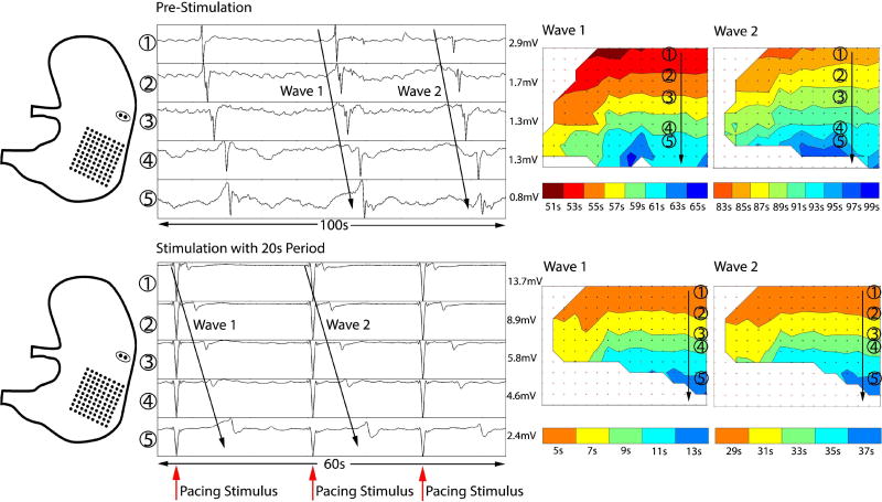
Fig. 11.
Validation of gastric pacing for modulating bradygastria, by simultaneous HR electrical slow wave mapping. Left: Diagram showing position of the stimulation leads and HR electrode mapping array for validation. Middle: Electrograms of slow wave activity before and after gastric pacing. Prior to stimulation, the slow wave frequency was bradygastric (period 30–40s). During simulation at period 20s, the frequency was increased to 3cpm. Right: HR propagation maps showing antegrade slow wave propagation over the anterior stomach before and after gastric pacing.

