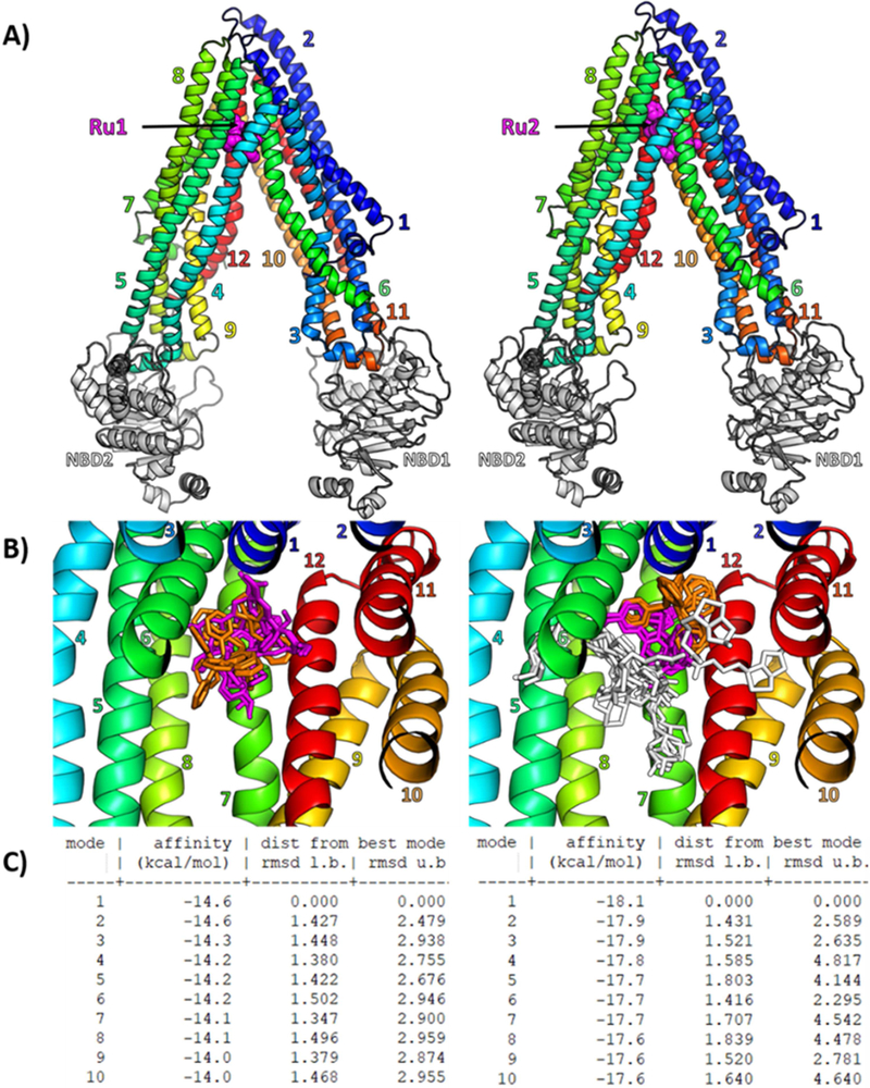
Figure 4.
(A) Cartoon representation of human P-gp 3D model, based on the mouse Pgp crystallographic structure (PDB 4Q9H), with the best docking pose of Ru1 and Ru2 computed with Autodock Vina in a gridbox containing 55 flexible residues forming the drug-binding pocket. Ru1 and Ru2 are shown as magenta spheres, transmembrane helices are numbered and colored from blue to red, Nucleotide Binding Domains (NBD) are colored in white. (B) Overview of the drug-binding pocket with all 10 flexible docking poses of Ru1 (left panel) and Ru2(right panel) shown as sticks with common core colored in magenta, common PPh3 in orange, and Ru2 additional R-group in white. (C) Docking affinities and RMSD are given for all 10 flexible docking poses (or “modes”) of Ru1 (left panel) and Ru2 (right panel).

