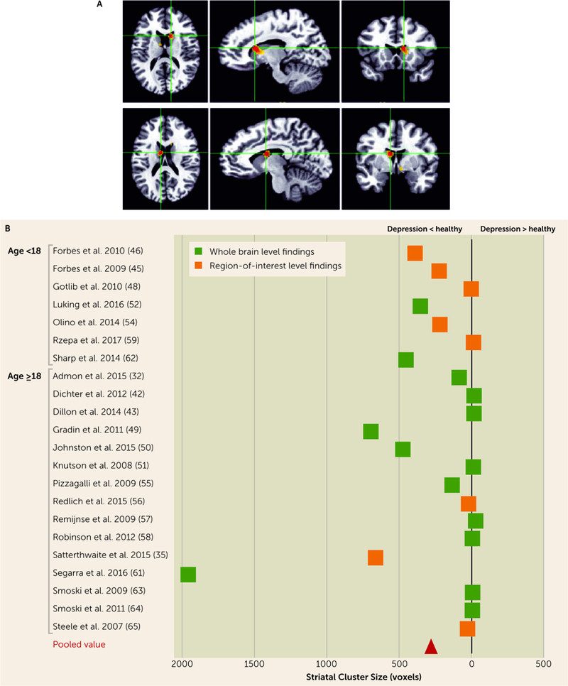
FIGURE 1. Alterations in Brain Activity During Reward Feedback, in Depressed Compared With Healthy Subjects: Meta-Analysis of fMRI Studiesa.

a Panel A depicts results across whole brain studies, presented as activation likelihood estimation maps, showing significantly decreased activation in the right caudate head and body (x=+12, y=+14, z=+14). Panel B lists the studies included in the meta-analyses of reward feedback contrast, broken down by age and type, along with the striatal cluster extent and direction of effect (increased versus decreased in depression). (The cluster value in the Johnston et al. study [50] was reported as 10,871 voxels combining several regions, and this is not reflected in its position in the graph because of space concerns.)
