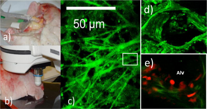
Figure 9. Real-time confocal microscopy of ex vivo lungs for reactive oxygen species (ROS) during reperfusion.

The ROS sensitive dye dihydrodifluorofluorescein (H2DFFDA, 10 μM) (Invitrogen, Molecular Probes, OR, USA) was added to the perfusate for 5-10 min prior to reperfusion. Images were acquired using a 10X lens on a Zeiss LSM 510 microscope. (a) Standard cannulation for continuous perfusion. (b) Stage preparation on the movable stage of Zeiss LSM 510 microscope for image acquisition. (c) Real-time imaging of ROS as detected by the green fluorescence of the oxidized H2DFFDAreactive oxygen species (d) A magnification of the box in c. (e). Polymorphonuclear Neutrophils (PMN) were labeled with red dye (CellTracker™ Red CMTPX, ThermoFisher Scientific, Waltham, MA, USA) and injected into the perfusate. After reperfusion, the lungs were washed and the PMN as detected by red fluorescence were observed sticking to the alveolar (Alv) and perivascular regions of the lungs.
