Fig. 3.
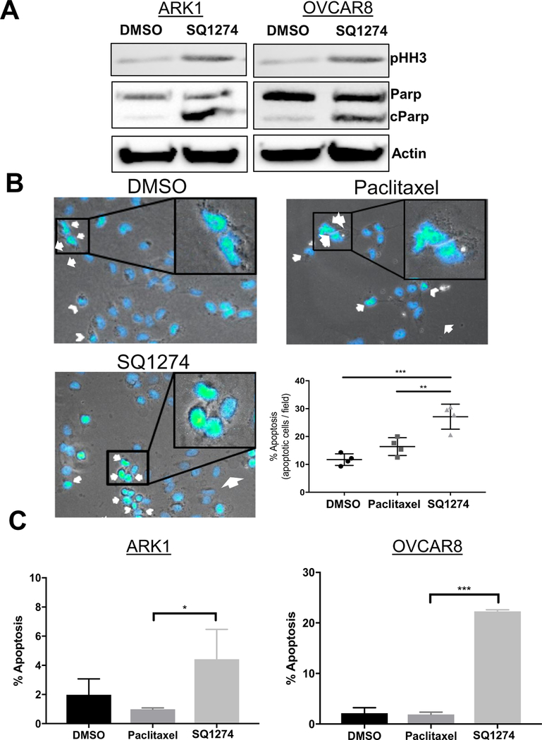
Mitotic arrest induces apoptosis. A) Western blot analysis of mitotic and apoptotic markers in ARK1 and OVCAR8 cells treated with DMSO or 2 nM SQ1274 for 24 h. B) Representative images and quantitation of apoptotic OVCAR8 cells treated with DMSO, paclitaxel, or SQ1274 detected by staining with DAPI and microscopic analysis of nuclear morphology. Arrow, abnormal DNA condensation; arrowhead, nuclear blebbing. **, P < 0.01; ***, P < 0.001. C) Quantitation of apoptotic ARK1 and OVCAR8 cells treated with DMSO, paclitaxel, or SQ1274 detected by staining with Annexin V/7-AAD and flow cytometric analysis. The mean apoptosis percentage ± SD are plotted, *, P < 0.05; ***, P < 0.001.

