
Figure 1.
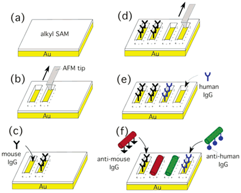
Schematic diagram to assemble anti-mouse IgG-coated nanotubes and anti-human IgG-coated nanotubes onto their antigen-patterned substrates via biological recognition. (a) Self-assembly of alkylthiol monolayers on Au substrates. (b) Shaving trenches on the alkylthiol SAM by using the AFM tip. (c) Deposition of mouse IgG on the shaved trenches. (d) Shaving another array of trenches on the alkylthiol SAM by using the AFM tip. (e) Deposition of human IgG on the shaved trenches. (f) Location-specific immobilization of Alexa Fluor 546-labeled anti-mouse IgG nanotubes onto the mouse IgG trenches and FITC-labeled anti-human IgG nanotubes onto the human IgG trenches via their biological recognition.