FIGURE 3.
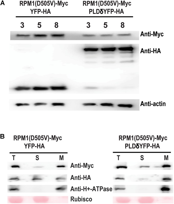
PLDδ reduces the protein level of RPM1(D505V) in N. benthamiana. (A) Protein level of RPM1(D505V). Est::RPM1(D505V)-Myc and 35S::PLDδ-YFP-HA were co-expressed in the leaves of N. benthamiana. RPM1(D505V) was induced with estradiol at 2 days after infiltration and the protein levels of RPM1(D505V)-Myc were detected at 3, 5, and 8 h after induction. 4 mM of LaCl3 was infiltrated into the leaves at 2 h before induction to block RPM1(D505V) induced cell death. Co-expression of YFP-HA and RPM1(D505V) was used as a control. Actin was used as a marker for protein equal loading. (B) PLDδ affects the membrane association of RPM1(D505V) in N. benthamiana. Samples were collected at 5 h after induction. The protein extraction solution (T) was separated into the cytosolic fraction (S) and the microsomal fraction (M) using centrifugation. The distribution of RPM1(D505V)-Myc in the two fractions were detected. The large subunit of Rubisco was used as a cytosolic protein marker. The plasma-membrane protein H+-ATPase was used as a microsomal protein marker. The RPM1(D505V)-Myc images were adjusted to the similar density to compare RPM1(D505V) levels distributed in the S fraction. The original western blots are available in Supplementary Materials.

