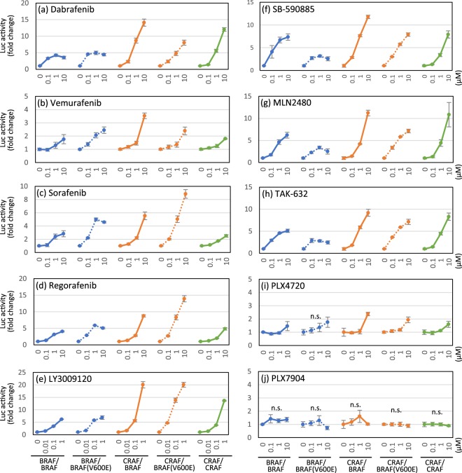
Figure 2.
Quantitative analysis of RAF dimer induction by RAF inhibitors using split luciferase probes. 293T cells were transiently co-transfected with a pair of RAF split luciferase probe plasmids (Table 1), stimulated with the indicated concentrations of RAF inhibitors for 2 hours, and luciferase activities were measured. Results are presented as fold increases compared with the luciferase activity of vehicle (0.1% DMSO)-treated cells (means ± SD, n = 3). n.s., not significant; otherwise P < 0.05 (one-way ANOVA). Statistical data are provided in Supplementary Table S3.

