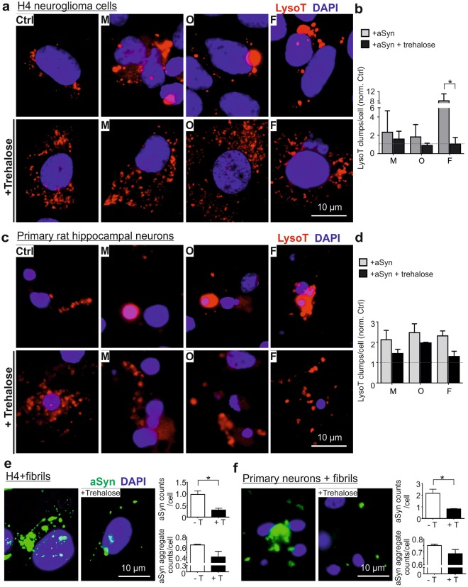
Figure 9.
Effects of trehalose on exogenous aSyn-induced lysosome dilation and aSyn accumulation. H4 cells (a,b) and primary neurons (c,d) were treated with trehalose prior to aSyn application (M: monomers; O: oligomers; and F: fibrils). (a,c) ICC analysis of lysosomes probed by LysoT. (b,d) The amount of larger LysoT+ clumps induced by aSyn decreases with trehalose-pretreatment. Quantification was conducted by normalizing the levels in exposed cells against untreated cells (Ctrl) (n = 3, Two-way ANOVA with Sidak’s multiple comparisons test). (e,f) ICC analysis of extracellular aSyn accumulated in fibril-exposed H4 cells (e) and primary neurons (f) with or without trehalose (+T or -T) pretreatment. Quantitative analysis of ICC images reveals that trehalose decreases the accumulation of extracellular aSyn by determining the counts of detectable aSyn signals/cell or by assessing the counts of larger aSyn aggregates (area >2 µm2)/cell (n = 3, unpaired t-test).

