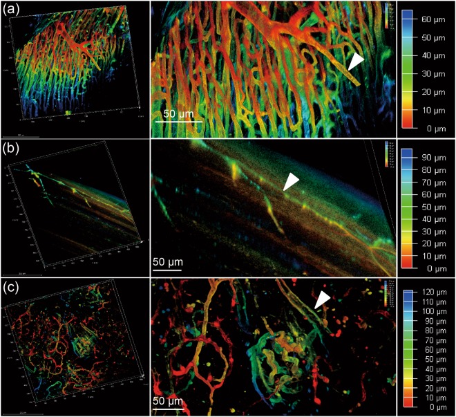
Figure 4.
Sections of heart, muscle and pancreas of a mouse are imaged using a 20x objective of a confocal microscope, after perfusion with MHI148-PEI and clearing with ECi. (a) Thin section from heart in 3D (left). Zoomed-in view shows arteries fully stained by MHI148-PEI. A scanned depth of 65 μm was used as indicated by the depth bar with color coding (right). (b) Thin muscle section from the leg of a mouse in 3D (left). Zoomed-in view shows capillary vessels with some branches (middle). A thin section of depth of about 90 μm was scanned as shown in the depth bar with color coding (right). (c) Thin section from pancreas (left). Zoomed-in view shows vessels in pancreas. A thin section of depth of about 120 μm was scanned as shown in the depth bar with color coding (right). The colored data in 3D are the structures and the black color is used for the background in each subfigure.

