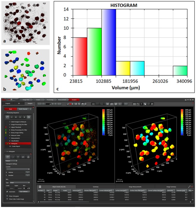
Figure 8.
Segmentation of glomeruli from mouse kidney perfused with MHI148-PEI and cleared with ECi using simple image processing pipeline: background removal using median filter, segmentation using Otsu thresholding in combination with binary morphological filters and 2-class classifier to filter only bigger glomeruli (class 1). The partially visible glomeruli and background objects were designated class 2. (a) Raw 3D data from small section of left mouse kidney. The raw data with specific settings is shown in Supplementary figure S13. (b) Segmented 3D data showing glomeruli using different group of colors based on the volume histogram shown in (c). (c) Volume histogram showing six groups where red color represents the group with smallest. (d) Leica LAS X software to create custom made analysis pipeline as shown by a sequence on left. The left and right images depict raw and segmented data (only class 2).

