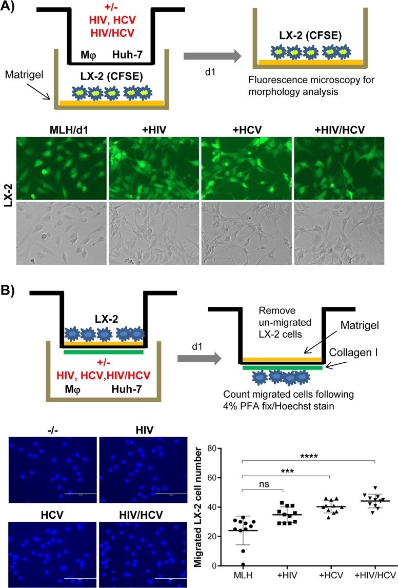
Figure 2.
HCV or HIV replication in MLH co-culture changed the morphological and invasive phenotypes of LX-2 cells. (A) Schematic of trans-well system separating LX-2 cells from Huh-7 and Mφ (top). Morphological characteristics of CFSE-labeled LX2 cells following MLH co-culture with or without HIV and HCV for 24 hr (bottom). (B) Schematic of LX2 cell invasion assay (top). Hoechst stained LX-2 cells detected at the bottom of trans-well insert following their migration (bottom left). The numbers of migrated LX-2 cells in three independent MLH co-cultures (bottom right). Asterisks indicate statistically significant difference measured by one-way ANOVA: ****p < 0.00005; ***p < 0.0005; ns, not significant.

