Figure 3.
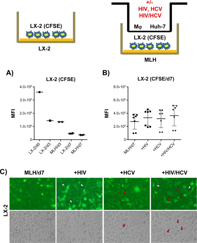
Mixed morphological changes in LX-2 cells induced by HCV/HIV co-infection of MLH co-cultures. (A) Proliferation of CFSE-labeled LX-2 cells during mono-culture and MLH co-culture during 7 day culture period determined by using flow cytometry. Day 0 (d0) indicates the time for initiating LX-2(CFSE) culture individually or under the condition of MLH co-culture, and the CFSE levels in LX2/d0 and MLH/d0 (data not shown) are same. (B) Effects of HCV and/or HIV replication in MLH co-culture on CFSE-labeled LX-2 cell proliferation determined by using FACS at day 7 of MLH co-culture. Results are from three independent experiments. (C) Morphology of LX-2 cells under MLH co-culture observed at day 7 by using a fluorescence and phase-contrast microscope. (C) LX-2 cell morphology under MLH co-culture at day 7 detected using a fluorescent and phase-contrast microscope.

