Fig. 1.
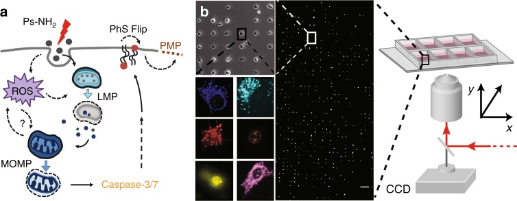
Nanoparticle-induced cell death studied by high-throughput single-cell time-lapse fluorescence microscopy. a Schematic illustration of the uptake and potential signaling pathways induced by exposure to 58 nm cationic polystyrene nanoparticles (PS-NH2 nanoparticles). Upon interaction with cells, nanoparticles can be directed into one of two interconnected pathways (i.e. LMP-dependent and MOMP-dependent) that trigger the activation of caspases 3/7, which ultimately results in the externalization of phosphatidylserine to the outer leaflet of the plasma membrane (PhS-Flip) and the permeabilization of the plasma membrane (PMP). b Schematic representation of the experimental single-cell platform. Cells are seeded on a micropatterned surface with one cell per square adhesion site (30 × 30 µm), and stained with a panel of fluorescence dyes to monitor key organelles and steps associated with cell death. Views of representative cells stained for mitochondria (blue), lysosomes (cyan), phosphatidylserine (red), nucleus (brown), caspase activation (yellow), and OxBurst (purple) are also shown. Scale bar: 200 µm

