Figure 4.
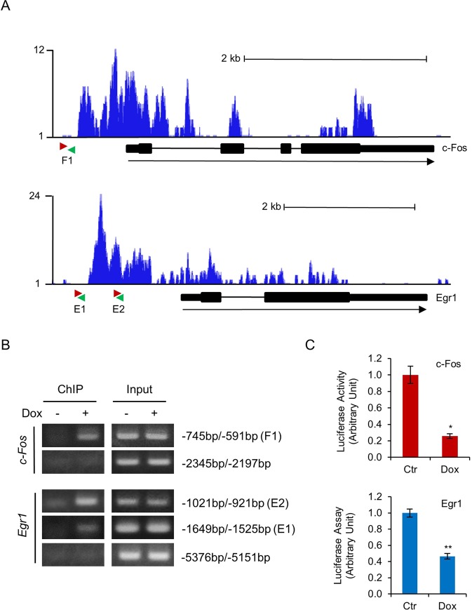
Gfi1 represses c-Fos and Egr-1 through direct binding to their promoters. (A) Gfi1 binding patterns at the c-Fos (upper panel) and Egr-1 (lower panel) based on the ChIP-seq data submitted by Möröy’s research group21. The pink and green arrows denote the forward and reverse primers, respectively, used to amplify the promoter regions at c-Fos (F1) and Egr-1 (E1 and E2) loci. (B) 32D/Y729F/Gfi1 cells were cultured with or without Dox for 24 hours. ChIP assays were carried out using the anti-mouse Gfi1 antibody. The indicated regions of c-Fos and Egr-1 promoters were amplified by PCR. (C) 32D/Y729F/Gfi1 cells were transfected with pGL3-basic vector containing c-Fos or Egr-1 promoter fragment and cultured in G-CSF with or without Dox. Luciferase activities were measured 24 hours later.

